- Use tools designed for urology
-
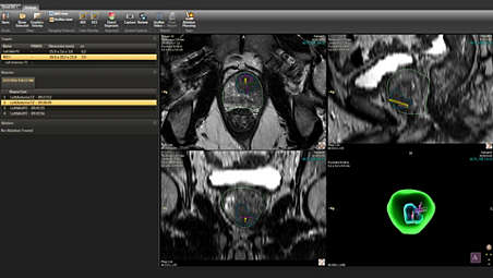
Use tools designed for urology
DynaCAD Urology works with DynaCAD Prostate, used in radiology, to provide urologists with an easy way to store, review and manage comprehensive diagnostic and therapeutic data. It provides viewing layouts and tools that are specifically dedicated for urological review. - Boost confidence for biopsies
-
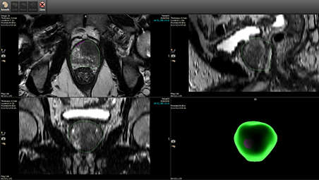
Boost confidence for biopsies
Once radiology has defined the prostate boundary and targets using DynaCAD Prostate, DynaCAD Urology displays the resulting information for urological review. Dedicated tools allow you to edit the prostate segmentation if needed, and add targets based on prior biopsy procedure core locations. This creates a ready-to-use plan for fusion-guided biopsy using Philips UroNav. - Seamless data export to external systems
-
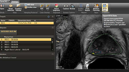
Seamless data export to external systems
DynaCAD Urology allows users to export unencrypted T2 MR images, gland and target segmentations and biopsy core locations to be used in external therapy planning and therapy systems. Exporting these from DynaCAD Urology streamlines the overall workflow by removing the need for Radiologists to segment the gland and targets separately for biopsy and therapy purposes. - Streamline your workflow
-
Streamline your workflow
An automatic exchange ensures that data moves quickly and reliably between the radiology and urology departments, bypassing possible delays such as when using discs or memory sticks. Secure network connectivity can also help enhance data integrity and safety – giving you the data you need for active surveillance, subsequent biopsy and therapy planning, and for follow-up.
Use tools designed for urology
Use tools designed for urology
Use tools designed for urology
Boost confidence for biopsies
Boost confidence for biopsies
Boost confidence for biopsies
Seamless data export to external systems
Seamless data export to external systems
Seamless data export to external systems
Streamline your workflow

Streamline your workflow
Streamline your workflow
- Use tools designed for urology
- Boost confidence for biopsies
- Seamless data export to external systems
- Streamline your workflow
- Use tools designed for urology
-
Use tools designed for urology
DynaCAD Urology works with DynaCAD Prostate, used in radiology, to provide urologists with an easy way to store, review and manage comprehensive diagnostic and therapeutic data. It provides viewing layouts and tools that are specifically dedicated for urological review. - Boost confidence for biopsies
-
Boost confidence for biopsies
Once radiology has defined the prostate boundary and targets using DynaCAD Prostate, DynaCAD Urology displays the resulting information for urological review. Dedicated tools allow you to edit the prostate segmentation if needed, and add targets based on prior biopsy procedure core locations. This creates a ready-to-use plan for fusion-guided biopsy using Philips UroNav. - Seamless data export to external systems
-
Seamless data export to external systems
DynaCAD Urology allows users to export unencrypted T2 MR images, gland and target segmentations and biopsy core locations to be used in external therapy planning and therapy systems. Exporting these from DynaCAD Urology streamlines the overall workflow by removing the need for Radiologists to segment the gland and targets separately for biopsy and therapy purposes. - Streamline your workflow
-
Streamline your workflow
An automatic exchange ensures that data moves quickly and reliably between the radiology and urology departments, bypassing possible delays such as when using discs or memory sticks. Secure network connectivity can also help enhance data integrity and safety – giving you the data you need for active surveillance, subsequent biopsy and therapy planning, and for follow-up.
Use tools designed for urology
Use tools designed for urology
Use tools designed for urology
Boost confidence for biopsies
Boost confidence for biopsies
Boost confidence for biopsies
Seamless data export to external systems
Seamless data export to external systems
Seamless data export to external systems
Streamline your workflow
Streamline your workflow
Streamline your workflow
Related products
Alternative products
-
UroNav
- MR/US guided fusion biopsy system
- Transperineal or transrectal options
- Targeted biopsy
View product
-
DynaCAD Prostate
- The advanced post-processing engine for a clear view
- Up to 12 customizable viewing options tailored to the user preferences.
- Automated segmentation to analyze lesions with volume, composition, and 3D views.
- customizable patient report templates with useful data of lesions
- Improves the performance and usability.
View product


-
UroNav
UroNav fuses pre-biopsy MR images of the prostate with ultrasound-guided biopsy images in real time, for excellent delineation of the prostate and suspicious lesions, as well as clear visualization of the biopsy needle path. Combining electromagnetic tracking and navigation with an onboard computer and a real-time imaging interface, UroNav brings precision targeting to your clinical practice in one easy-to-use, mobile workstation.
View product
-
DynaCAD Prostate
Offers a comprehensive set of tools for real-time analysis, review, and reporting of multi-parametric, multi-vendor(3) MRI studies. Enhances productivity by transferring images directly from the MRI to DynaCAD and utilizing its robust, automatic post-processing tools and display the results in customized hanging protocols for analysis and reporting.
View product
- DynaCAD Urology and DynaCAD Prostate are modules of the DynaCAD product.