
FieldStrength MRI magazine
User experiences - July 2015
Used in most of its MSK exams, mDIXON TSE shines at CHRU
Lille
The musculoskeletal imaging department at Lille University Hospital (CHRU, Lille, France) manages to get more information in the same time slot after integrating mDIXON TSE in many exams. mDIXON can provide four different contrasts from a single acquisition and its fat suppression is robust, helping us reduce additional scans.
The Hospital’s musculoskeletal imaging department, led by Prof. Anne Cotten, scans patients referred from various departments including emergency, orthopedics, neurosurgery and rheumatology using its Ingenia 3.0T system. According to Guillaume Lefebvre, MD, the MRI team has recently changed its way of working by integrating mDIXON TSE into most MR exams of peripheral joints and spine. Using mDIXON TSE has contributed to detailed diagnoses and diagnostic confidence, he says.

Guillaume Lefebvre, MD is an MSK Radiologist at Lille University Hospital (Pr. A. Cotten ‘s Department). Dr. Lefebvre is a member of French Society of Radiology (SFR), Society of Musculoskeletal Imaging (SIMS), European Society of Musculoskeletal Radiology (ESSR), European Society of Radiology (ESR), and Radiological Society of North America (RSNA).
A way to get more information within the available time
“In MSK imaging, we generally need T1-, T2-, PD-weighted, and sometimes post-contrast sequences, with or without fat suppression, in different orientations. The goal is to have enough information to see abnormalities in signal and morphology, but we can't fit all orientations, all weightings and all contrasts into an exam, so we must make a choice.” “With previous exam protocols, we sometimes missed information because we didn’t choose the right acquisitions, with or without fat suppression for example. Adding sequences in our exam could help, but lengthens the examination time and thus reduces the availability of MRI. A long exam is also a source of patient motion, which degrades the image quality.” “mDIXON TSE helped us find a solution here, as it provides four different contrasts in just one acquisition – an important reason why we prefer using it. mDIXON TSE is now added to most joint MRI protocols at our hospital.”
Bone assessment with confidence
“For bone assessment near joints, mDIXON TSE provides the visualization and multiple contrasts to describe abnormalities within a limited number of acquisitions. Bone marrow signal abnormalities are common MRI findings that can represent various underlying causes, from normal variance to malignancy. So, it is important for us to notice and characterize these findings. With different contrasts, both with and without fat signal as mDIXON TSE efficiently provides, we can make a confident diagnosis.” “Other examples in bone are the signal description of a necrotic fragment in osteonecrosis, the signal description of tumoral matrix that has different components (necrosis, hemorrhage, cartilage, bone formation). These are all possible thanks to in-phase and water images from a single mDIXON acquisition.”
“We prefer using mDIXON TSE, which provides four different contrasts in the same acquisition.”
“Radiologists no longer need to choose between fat suppression or not.”
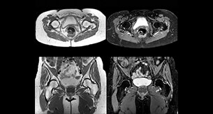
Add fat suppressed imaging without time penalty for peripheral joints
“In peripheral joints, mDIXON TSE imaging aids in diagnosing injuries in ligaments or tendons, for imaging degenerative and inflammatory pathologies such as osteoarthritis and rheumatologic disorders and for oncological exploration.” “For tendon and ligament assessment around knee, ankle, hip and elbow, mDIXON TSE contributes to diagnostic confidence thanks to having images both with and without fat suppression – and without time penalty. This is possible because 2-point mDIXON is faster than the common 3-point Dixon method. It can also increase efficiency as it helps avoid having to add scans during the exam.”
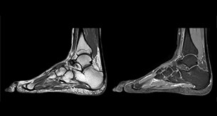
Amazing quality of fat suppression
“In peripheral joints, we get good image quality in difficult areas with mDIXON TSE.
Fat suppressed images appear homogeneous over the entire image, even with large coverage at 3.0T – for instance in scapular or hip girdles – or in the bearing areas or around metal prostheses, where fat suppression is often deficient with STIR or spectral fat suppression, causing diagnostic difficulties. If a diagnostic image is right the first time, we don’t need to repeat or add a sequence.” “mDIXON TSE sequences allow simultaneous characterization of morphological changes from the in-phase T2-weighted images and visualization of edematous changes, thanks to the water T2-weighted images from the same acquisition. Anatomical and morphological considerations could be a partial or complete ligament tear, a bony avulsion or hematoma.” “For soft tissue assessment mDIXON brings similar benefits. For example in one T2-weighted mDIXON TSE acquisition, having the multiple contrasts helps us assess abnormalities in peripheral nerves fascicles, which may be due to anatomical or inflammatory changes..”
“Fat suppressed images appear homogeneous over the entire image, even at 3.0T.”
Improving efficiency in musculoskeletal MRI
Dr. Lefebvre appreciates the efficiency of mDIXON TSE. “Using mDIXON TSE we can reduce the number of sequences scanned, without reducing the number of contrast types provided. In addition, we also value the reduction we see in repeats because of artifacts (as frequently found in other fat suppressed sequences as SPAIR and SPIR), making the acquisition non-diagnostic. Imaging right the first time is the most efficient way for us, and also for the patient.” “Using mDIXON TSE gives us homogeneous fat suppression in a reduced scan time, as well as good correlation between different images. In addition, we have a direct match between different contrasts because they are obtained from the same acquisition.”
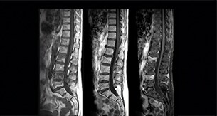
Spine scans must be fast and complete
“In our spine cases, we use mDIXON TSE for patients with degenerative and inflammatory spine issues, vertebral fractures and vertebral and paravertebral tumor characterization,” says Dr. Lefebvre. “It provides, in a single acquisition, different contrasts so we can both visualize and characterize spinal, focal or diffuse spine lesions.”
“mDIXON TSE makes real a difference in cases of acute and traumatic spinal injuries, where it allows us to reduce the scan time by only using a limited number of sequences. For patients in pain, who come from the emergency unit and need surgery, for example, MRI must be very fast. With a single T2-weighted acquisition we can assess spinal cord, vertebral disc and ligament wholeness with in-phase images, as well as trabecular fracture and edematous changes with water images.” Dr. Lefebvre cites other frequent examples of spinal diseases that benefit from the mDIXON technique. “In cases of vertebral disc herniation or degenerative discs, for example, we don’t have to choose between fat or no fat images. We can assess morphological changes in the endplate with in-phase images from T2-weighted sequences and in the water image from the same acquisition we will see inflammatory changes into endplates.”
Improvements in the way of working
Dr. Lefebvre says, “Our radiologists no longer need to choose between fat suppression or not during the examination; they have both. In addition they appreciate mDIXON TSE for exceptional image quality and homogeneous fat suppression. Using mDIXON TSE can even help make diagnoses that were difficult or not possible with our older fat suppression methods. In joints, for example, we get better image quality in difficult areas where the fat removal used to be usually deficient.” “Our referring physicians also appreciate the high quality images for confident diagnosis and optimal care.
For technologists, using mDIXON TSE helps reduce errors related to the positioning of fat saturation bands, normally used with fat sat acquisitions. Our department managers like to see short scan times, which lead to increased MRI availability.
Easy to use and highly recommended
Overall, Dr. Lefebvre reports, “I would definitely recommend mDIXON TSE for MSK imaging. It’s an easy way to get a major improvement of an MRI examination, with simultaneous images for visualization and characterization without time penalty.”
Reference
1. Del Grande F, Santini F, Herzka DA, Aro MR, Dean CW, Gold GE, et al. Fat-suppression techniques for 3-T MR imaging of the musculoskeletal system. Radiographics, Feb 2014;34(1):217-233.
“Using mDIXON gives us homogeneous fat suppression in a reduced scan time.”
What Dr. Lefebvre likes about mDIXON TSE
Dr. Lefebvre’s tips for mDIXON TSE in MSK
* Metal implants are a contraindication for MRI, unless the MR compatibility for the implant is stated by the implant manufacturer. Results from case studies are not predictive of results in other cases. Results in other cases may vary. Results obtained by facilities described in this issue may not be typical for all facilities.

Clinical cases
“We can both visualize and characterize spinal, focal or diffuse spine lesions.”
Related information
More from FieldStrength